Laser Scanning Confocal Microscope Maximum Magnification

What Is Confocal Laser Scanning Microscopy

Confocal Microscopy Introduction Olympus Life Science

Laser Scanning Confocal Microscopy Lscm Reflexion Mode Images At Two Download Scientific Diagram

Biophotonics Lecture 16 November Fourier Plane Point Object Image F F F F Magnification M 1 Angles Sin Sin Magnification Ppt Download
How Does A Confocal Microscope Work
Confocal Microscopy Confocal Microscope Scanning Systems Olympus Life Science
The confocal laser scanning microscope s aim was not to further increase magnification but to make clearer.
Laser scanning confocal microscope maximum magnification. The laser scans across the object and an image is built up pixel by pixel on a screen. Another drawback is that duv microscopes do not have the capability for surface topography 3d imaging. The confocal laser scanning microscope clsm is a microscope which focuses only on a single focal plane and the unfocused plane will not be visualized. The scanning electron microscope has a magnification range from 15x to 200 000x reached in 25 steps and a resolution of 5.
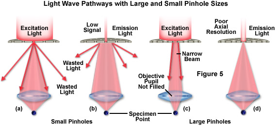
In the past the traditional laser microscope excited the whole thickness of the sample resulting in saturated blurry images and sometimes visualizing false colocalization images. What is the maximum magnification of the scanning electron microscope. This resulted in a costly microscope system with magnification limited to 1000x. Laser scanning confocal microscopy laser scanning confocal microscopes employ a pair of pinhole apertures to limit the specimen focal plane to a confined volume approximately a micron in size.
The laser scanning microscope uses a scanning design called beam scanning where the laser image path is scanned in a raster pattern on the surface of the sample. Confocal microscopy most frequently confocal laser scanning microscopy clsm or laser confocal scanning microscopy lcsm is an optical imaging technique for increasing optical resolution and contrast of a micrograph by means of using a spatial pinhole to block out of focus light in image formation.

Confocal Laser Scanning Microscopy An Overview Sciencedirect Topics

A Practical Guide For Fluorescent Confocal Microscopy The Marder Lab

Laser Scanning Confocal Microscope Tailored Tutors

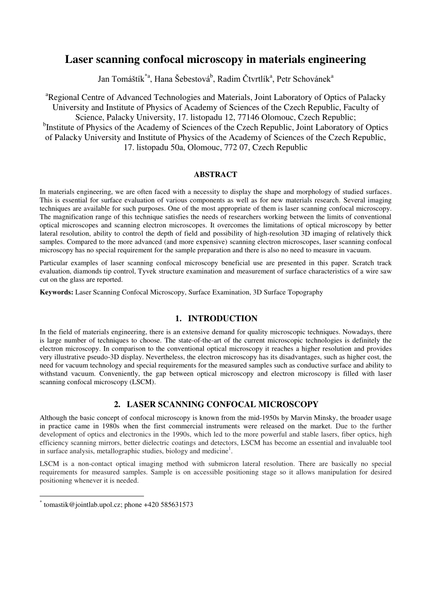
Nikon A1si Laser Scanning Confocal Microscope Washington University Biology Imaging Facility

Introduction To Spinning Disk Confocal Microscopy

33 Laser Scanning Confocal Microscopy And Laser Microdissection Musculoskeletal Key
Zeiss Microscopy Online Campus Introduction To Spinning Disk Microscopy
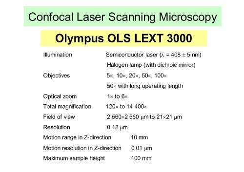
Confocal Laser Scanning Microscopy Olympus Ols Lext 3000
Confocal Microscopes Microscope Imaging Network

Confocal Laser Scanning Microscope An Overview Sciencedirect Topics

Laser Scanning Confocal Microscopy Of Scots Pine Seedlings Colonized By Download Scientific Diagram
Homework 2 Confocal Microscopy 04 05 12

Confocal Microscopy An Overview Sciencedirect Topics
Pdf Laser Scanning Confocal Microscopy In Materials Engineering

Pdf Advanced Microscopy Laser Scanning Confocal Microscopy

Confocal Microscopy For Real Time Detection Of Oral Cavity Neoplasia Clinical Cancer Research

Inverted Zeiss Lsm880 Laser Scanning Confocal Microscope With Airyscan Cell Sciences Imaging Facility Csif

Advances In Bioscience Education Summer Workshop Ppt Video Online Download
Https Encrypted Tbn0 Gstatic Com Images Q Tbn 3aand9gcr3fysoxor5w4y0kayjtt5nby84 Yhi3vdxn3rx2 E Usqp Cau

Laser Scanning Confocal Microscopy History Applications And Related Optical Sectioning Techniques Radiology Key
Fv3000 Confocal Laser Scanning Microscope From Olympus Life Science Solutions Get Quote Rfq Price Or Buy
Applied Sciences Free Full Text Contrast Enhancement For Topographic Imaging In Confocal Laser Scanning Microscopy Html

Olympus Confocal Microscope Lext Ols3100 Tel Aviv University Center For Nanoscience And Nanotechnology
Confocal Microscopy Dinesh
Pdf Surface Roughness Determination Using Laser Scanning Confocal Microscope Zeiss Lsm 700

Molecular Expressions Microscopy Primer Virtual Microscopy Laser Scanning Confocal Microscopy
Laser Scanning Confocal Microscope Cm Olympus Fluoview Fv1000 Download Scientific Diagram
Pdf A 3d Imaging And Visualization Workflow Using Confocal Microscopy And Advanced Image Processing For Brachyuran Crab Larvae

Pdf Using Confocal Laser Scanning Microscopy Scanning Electron Microscopy And Phase Contrast Light Microscopy To Examine Marine Biofilms
Olympus Launches Two Types Of Upright Fv3000 Confocal Laser Scanning Microscope 2017 News Olympus

Laser Scanning Confocal Microscopy Nikon S Microscopyu
Confocal Microscopy Signal To Noise Considerations Olympus Life Science

Inverted Zeiss Lsm 780 Multiphoton Laser Scanning Confocal Microscope Cell Sciences Imaging Facility Csif

Introduction To Microscopy Duke Light Microscopy Core Facility

Focal Wars Widefield Vs Confocal Biocompare The Buyer S Guide For Life Scientists
.jpg)
A 3 In 1 Measurement Technology That Can Make The Difference

Scanning Optical Microscopy Emmi Laser Scan Obirch Som

Pdf Application Of Confocal Laser Scanning Microscopy In Dentistry

Laser Scanning Confocal Microscopy Java Tutorial Olympus Life Science

Pdf Confocal Microscopy Applications In Materials Science

The Integrated High Resolution Reflection Mode Photoacoustic And Fluorescence Confocal Microscopy Sciencedirect

Modern Laser Scanning Confocal Microscopy Bayguinov 2018 Current Protocols In Cytometry Wiley Online Library