Laser Scanning Microscope Max Magnification

The confocal laser scanning microscope s aim was not to further increase magnification but to make clearer.
Laser scanning microscope max magnification. Magnification image contrast laser intensity detector sensitivity flip rotate image line scan and area scan modes are user selectable includes laser scanning confocal microscope lscm controller controller cables galvanometer data communication power interlock and connector kit. Clsm combines high resolution optical imaging with depth selectivity which allows us to do optical sectioning. A transmission electron microscope tem produces a 2d image of a thin sample and has a maximum resolution of 500000. Capturing multiple two dimensional images at different depths in a sample enables the.
Confocal microscopy most frequently confocal laser scanning microscopy clsm or laser confocal scanning microscopy lcsm is an optical imaging technique for increasing optical resolution and contrast of a micrograph by means of using a spatial pinhole to block out of focus light in image formation. Relatively thick specimens can be imaged in successive volumes by acquiring a series of sections along the optical z axis of the microscope. Separating light waves with lasers and with. This feature is termed the zoom factor and is usually employed to adjust the image spatial resolution by altering the scanning laser sampling period.
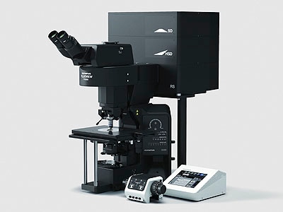
A scanning electron microscope sem produces a 3d image of a sample by bouncing electons off and dectecting them at multiple detectors. A confocal laser scanning microscope or also known as laser scanning confocal microscope is used to obtain high resolution images and generate 3d reconstructions through direct optical sectioning to provide clear images from a range of depths of thicker specimens for nanometer level imaging and measurement. In the confocal laser scanning microscope the highest frequency to be sampled f is imposed by the optical system and for a particular resolution specification. In some cases specimens should be sampled at more than 2 3 times the highest information frequency to allow for the possibility that the highest frequency was misjudged.
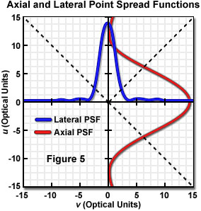
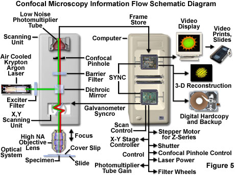
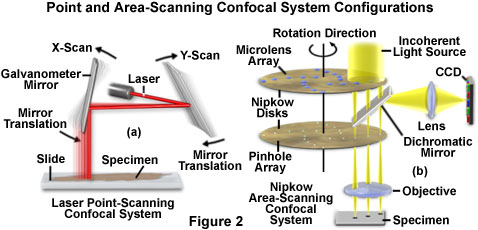
Laser scanning confocal microscopy laser scanning confocal microscopes employ a pair of pinhole apertures to limit the specimen focal plane to a confined volume approximately a micron in size. This means that we can view visual sections of tiny structures that. The laser scans across the object and an image is built up pixel by pixel on a screen. Fluorescent microscopy not only makes our images look good it also allows us to gain a better understanding of cells structures and tissue.
Additional advantages of scanning confocal microscopy include the ability to adjust magnification electronically by varying the area scanned by the laser without having to change objectives. The confocal laser scanning microscopes enable researchers to create detailed 3d pictures of cell organelles and to examine live cells through incubation systems that facilitate the study of cellular changes over time.









.jpg?rev=33A9)


